∞ generated and posted on 2016.01.14 ∞
In light microscopy this is where the light comes from that you see when viewing a specimen.
In modern light microscopes the light source is a light bulb. The detection instrument is the human eye. Cameras and, more recently, charge-coupled devices can be employed as well, particularly given microscopes designed to accommodate these devices (i.e, trinocular microscopes).
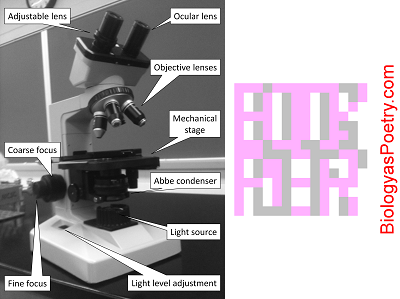
The pathway of light through a compound microscope is: light source → condenser → iris diaphragm → stage → object/specimen → objective lens → ocular lens → eye or camera. See equivalently, illuminator.

Figure legend: The light source can be found at the bottom of this microscope, center, towards the front, beneath the indicated Abbe condenser. Note as well the light level adjustment, which here is found to the side. A separate on-off switch may be present as well.
There will be an on and off switch and/or a means by which one can control the amount of light that is given off. Typically one employs the maximum amount of light available though those who are light sensitive may chose to reduce the amount of light coming from the light source. One of the first things to check when trying to debug microscopy problems is whether or not the light source is turned sufficiently high.